www.Smoothmap.org |
||||
last updated: 23 April, 2016 |
||||
www.Smoothmap.org |
||||
last updated: 23 April, 2016 |
||||
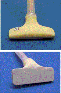 |
Small Intestine Electrode. 16 electrodes (3 x 4 + 1; 5-7 mm inter-electrode distance) with electrode tips flush with the electrode array. The electrode array was developed and built for mapping the small intestine by the group of Alan Bradshaw and Bill Richards at Vanderbilt University, Nashville in close co-operation with Andrew Pullan and Leo Cheng at Auckland University, NZ. The electrode is currently used for recording slow wave activities in small intestine in humans and in pigs. The array was developed and constructed by Atiq Al Wahab (2006/2007). It includes several novelties such as the possibility of bending the cable in different shapes and curves. A similar electrode array, containing 32 individual electrodes, was also built for the group at Auckland University, New Zealand (Andrew Pullan, Leo Cheng and John Windsor). |
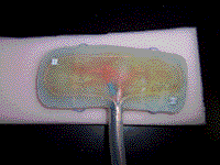 |
Pig Stomach Electrodes. This is a slightly curved mapping electrode consisting of 48 electrodes, in four rows of 12 each with an inter-electrode distance of 9 mm. To be used for recording slow waves on the stomach in anaesthetized open-abdominal model of the pig. Made by Atiq Al Wahab in 2007/2008. In the magnified picture, the left photo shows the individual components of the assembly with the 48 soldered electrode wires, a sleeved copper loop for stability and several (blue) plastic loops. The middle photo shows the finished assembly (mixture of epoxy and silicon glue) with several suture loops visible. The third photo shows the whole system with the electrode array, a 3 meter shielded connecting bundle and 48 separate female connectors. This platform was built for the group of Alan Bradshaw and Bill Richards at the Vanderbilt University, Nashville. |
 |
|
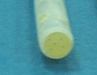 |
Laparoscopic Electrodes. Five closely-spaced teflon-coated silver leads were constructed in a 6 mm (o.d.), 30 cm long, Teflon tube, filled and fixed with epoxy resin. The five individual silver wires sere soldered to five Teflon-coated multi-strand stainless steel wire, sleeved in a silicone tube (2 meters length) and soldered to bayonet jack and socket connector. Several laparoscopic electrode sets (also described as "wands") have been built and are being used by the group in Auckland (Pullan, Cheng, O'Grady, Du, Egbuji and Windsor). This was again made by Athiq Wahab (2008). |
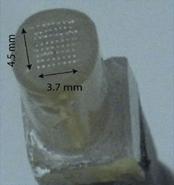 |
|
||
|
Sinus Node Mapping Electrode:
Close up of the electrode assembly head to show the 8x8 electrode configuration. The tips are 120 micron diameter and approximately 0.3-0.5 mm apart from each other. |
Overview of the whole assembly with the electrode head at the left top and the shielded 64-connecting wires at the bottom. The whole assembly has been right-angled for easy placement in the experimental set-up. |
||
 |
 |
||
|
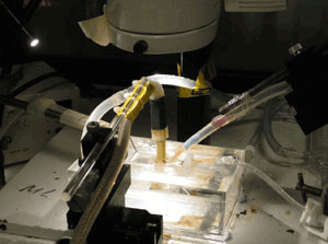
The tissue bath (Perspex) consists of a heated box with in the middle a rectangular depression for holding and superfusing the isolated SA-preparation. The mapping electrode is seen here in position, attached to a manipulator. To the right, the indifferent electrodes with their tips close to the preparation. |
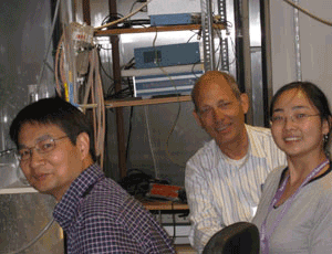
The SA-mapping team: Dr Ming Lei (front left), Dr Jing Jing Wu (front right) and myself (back). July 2008: Cardiovascular Research Group; Manchester University, UK. This electrode was also built by Athiq Wahab (Spring 2008). |
||
|
Second Small Intestine Electrode
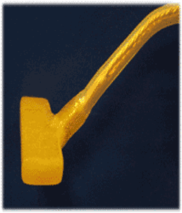
A 16 electrode array with electrode tips flush with the electrode base. It This is essentially the same type as the one at the top of this page. However, this array was built without a PVC base, using only epoxy as the outer layer, which improves on the issue of sterility and reliability.
The electrode array was developed and built for mapping the small intestine by the group of Alan Bradshaw and Bill Richards at Vanderbilt University, Nashville The array was developed and constructed by Atiq Al Wahab (2008). |
|
© W. Lammers 2001-2016
|
||